Home » Without Label » Chest Muscle Anatomy Diagram - The 5 Best Compound Chest Exercises You Can Do At Home ... : Neck and shoulder muscles diagram neck shoulder muscle anatomy shoulder muscle anatomy diagram anatomy.
Chest Muscle Anatomy Diagram - The 5 Best Compound Chest Exercises You Can Do At Home ... : Neck and shoulder muscles diagram neck shoulder muscle anatomy shoulder muscle anatomy diagram anatomy.
Chest Muscle Anatomy Diagram - The 5 Best Compound Chest Exercises You Can Do At Home ... : Neck and shoulder muscles diagram neck shoulder muscle anatomy shoulder muscle anatomy diagram anatomy.. The word sternum originates from the ancient greek word 'sternon', meaning chest. Computed tomography (ct) of the chest can detect pathology that may not show up on a conventional chest radiograph (1). Barbells are great for developing overall strength in your pressing muscles. Make your life and learning a lot easier by using kenhub's muscle anatomy reference. Chest muscle anatomy diagram :
Plus, how to target each to make them bigger and stronger. Related posts of chest muscle diagram muscle gross anatomy. Chest muscle anatomy diagram : Build your upper body with. Neck and shoulder muscles diagram the superficial back muscles attachments actions teachmeanatomy
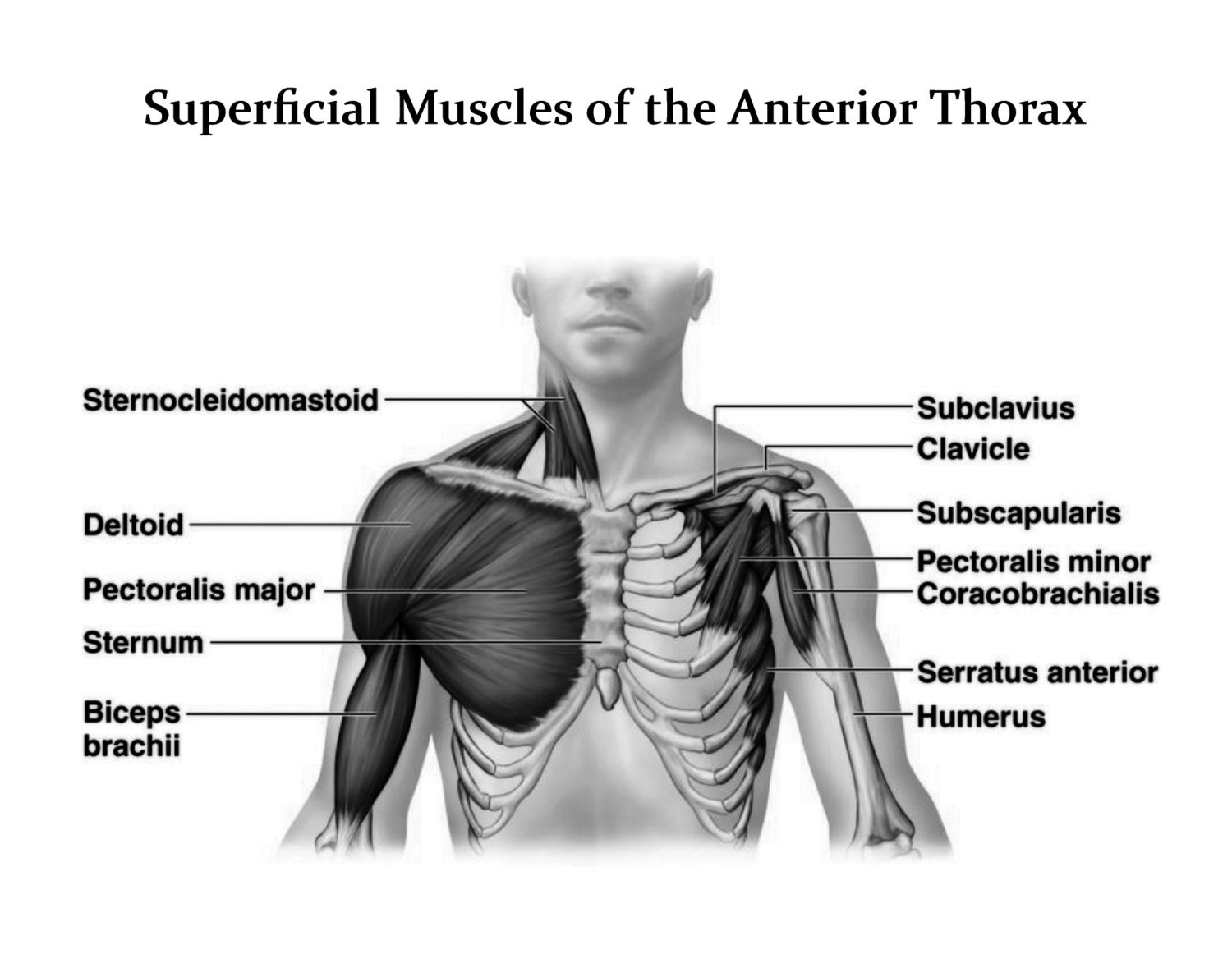
Muscles of the Chest - Muscles of the Anterior Thorax ... from img0.etsystatic.com Fill in your note sheet by clicking on the number and naming each muscle. The pectoralis major, the pectoralis minor, and the serratus anterior. Female chest muscle anatomy diagram / female chest muscles anatomy diagram function body maps. Several muscles that move the arms, head, and neck have their origins on the sternum. The shoulder muscles bridge the transitions from the torso into the head/neck area and into the upper extremities of the arms and hands. It also protects several vital organs of the chest, such as the heart, aorta, vena cava, and thymus gland that are located just deep to the sternum. It forms the bulk of the chest area and can be easily seen on the. The major muscle in the chest is the pectoralis major.
Anatomy and physiology of the muscles of the chest.
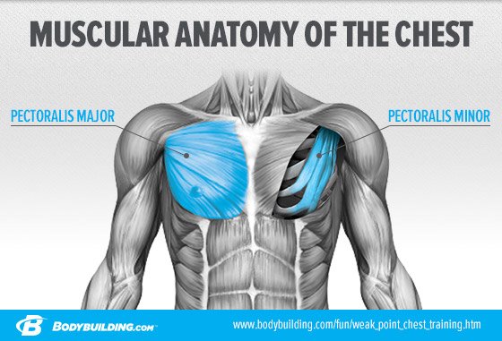
Female chest muscle anatomy diagram / female chest muscles anatomy diagram function body maps. See chest anatomy stock video clips. Learn about each of these muscles, their locations, functional anatomy and exercises for them. Fill in your note sheet by clicking on the number and naming each muscle. I know anatomy guides aren't something i've posted before, but i figured i'd put this out there for whoever could find it helpful. Leg muscles diagram unlabeled : Find out more about the individual muscles within the chest anatomy by clicking their respective. Computed tomography (ct) of the chest can detect pathology that may not show up on a conventional chest radiograph (1). The major muscle in the chest is the pectoralis major. Learn about each of these muscles, their locations, functional anatomy and exercises for them. System respiratory respiratory organs of human body digestive and respiratory system medical chest internal structure of human body medicine body lungs biology intestines stomach anatomy torso human internal. Make your life and learning a lot easier by using kenhub's muscle anatomy reference. Of the two chest muscles, the pectoralis major (a.k.a.
Muscular male chest vector icon. Neck and shoulder muscles diagram neck shoulder muscle anatomy shoulder muscle anatomy diagram anatomy. Labeled anatomy chart of neck and shoulder muscles on black background labeled human anatomy diagram of man's neck and shoulder muscles in an anterior view on a black background. Leg muscles diagram unlabeled : Start with a pair of dumbbells extended above your chest.
Objectives-2, BIO 2310, Coloring Exercise | Clare Hays ... from sites.msudenver.edu Chest muscles, chest muscle diagram. Make your life and learning a lot easier by using kenhub's muscle anatomy reference. Muscular male chest vector icon. The pec major) is the one that commands the most real estate. For that reason, and because of the dexterity of the shoulder joint itself, the musculature of the shoulder is. Several muscles that move the arms, head, and neck have their origins on the sternum. Computed tomography (ct) of the chest can detect pathology that may not show up on a conventional chest radiograph (1). The sternum is located along the body's midline in the anterior thoracic region just deep to the skin.
Learn about each of these muscles, their locations, functional anatomy and exercises for them.
Here, we break down the anatomy of your chest muscles. Wiring rj45 ends wiring schematic symbols chart wiring house diagram wiring schematics a wiring diagram for a 2005 chevy cobalt ignition coil pack wiring meter. Labeled anatomy chart of neck and shoulder muscles on black background labeled human anatomy diagram of man's neck and shoulder muscles in an anterior view on a black background. 'partition'), is a sheet of internal skeletal muscle in humans and other mammals that extends across the bottom of the thoracic cavity.the diaphragm separates the thoracic cavity, containing the heart and lungs, from the abdominal cavity and performs an important function in. Make your life and learning a lot easier by using kenhub's muscle anatomy reference. Several muscles that move the arms, head, and neck have their origins on the sternum. This page provides an overview of the chest muscle group. 2, all parts of the pm may be more or less separable. The gastrocnemius and soleus muscles taper and merge at the base of the calf muscle. Female chest muscle anatomy diagram / female chest muscles anatomy diagram function body maps. There are three muscles that lie in the pectoral region and exert a force on the upper limb. It is a flat bone that articulates with the clavicle and the costal cartilages of the upper 7 ribs (true ribs), while the 8th, 9th and 10th ribs (false ribs) are indirectly attached with sternum via costal cartilage of the ribs above. The major muscle in the chest is the pectoralis major.
Computed tomography (ct) of the chest can detect pathology that may not show up on a conventional chest radiograph (1). Learn about each of these muscles, their locations, functional anatomy and exercises for them. Of the two chest muscles, the pectoralis major (a.k.a. Learn about each of these muscles, their locations, functional anatomy and exercises for them. The muscles of the chest and upper back occupy the thoracic region of the body inferior to the neck and superior to the abdominal region and include the muscles of the shoulders.
Wimpy Chest No More: 3 Chest Routines For Massive Growth! from www.bodybuilding.com Neck and shoulder muscles diagram the superficial back muscles attachments actions teachmeanatomy Anterolateral trunk muscles diagram the anterior trunk muscles cover the anterolateral part of the trunk. Related posts of muscles of the arm and forearm diagram piriformis muscle anato… august 15, 2021. Here, we break down the anatomy of your chest muscles. The dominant muscle in the upper chest is the pectoralis major. / chest muscles, chest muscle diagram. Muscle gross anatomy 12 photos of the muscle gross anatomy gross anatomy of cardiac muscle, gross anatomy of skeletal muscle worksheet, gross muscle anatomy test, muscle gross anatomy quiz, muscular system gross anatomy chapter 10, human muscles, gross anatomy of cardiac muscle, gross anatomy of skeletal muscle worksheet, gross. Educational video describing the muscle anatomy and function of the pectoralis muscles.the human chest consists of two pectoral muscles,.
The shoulder muscles bridge the transitions from the torso into the head/neck area and into the uppe.
Neck and shoulder muscles diagram neck shoulder muscle anatomy shoulder muscle anatomy diagram anatomy. Here, we break down the anatomy of your chest muscles. Of the two chest muscles, the pectoralis major (a.k.a. Labeled anatomy chart of neck and shoulder muscles on black background labeled human anatomy diagram of man's neck and shoulder muscles in an anterior view on a black background. Barbells are great for developing overall strength in your pressing muscles. Learn about each of these muscles, their locations, functional anatomy and exercises for them. It is a flat bone that articulates with the clavicle and the costal cartilages of the upper 7 ribs (true ribs), while the 8th, 9th and 10th ribs (false ribs) are indirectly attached with sternum via costal cartilage of the ribs above. Several muscles that move the arms, head, and neck have their origins on the sternum. System respiratory respiratory organs of human body digestive and respiratory system medical chest internal structure of human body medicine body lungs biology intestines stomach anatomy torso human internal. It also protects several vital organs of the chest, such as the heart, aorta, vena cava, and thymus gland that are located just deep to the sternum. The major muscle in the chest is the pectoralis major. Leg muscles diagram unlabeled : 2, all parts of the pm may be more or less separable.